

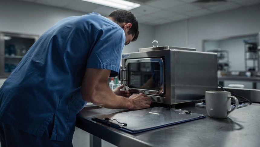
A preserved eyeball sits in a machine designed to keep it alive, tethered to tubes and sensors, waiting for a surgeon’s hands.
For decades, whole-eye transplantation has remained one of medicine’s most stubborn failures. The surgery itself is technically possible. The real problem is time: human eyes begin degrading the moment they leave a donor’s body, and that window of viability has always been too narrow to make transplants reliable enough to justify the risks. But a new device is changing that calculus entirely, potentially unlocking the first genuinely successful pathway to restoring sight to people who have lost it.
- The Viability Barrier: The retina begins to suffer irreversible damage within minutes of losing blood flow, making donor eye preservation one of transplant medicine’s hardest unsolved problems.
- The Machine’s Breakthrough: A new ex vivo perfusion device can keep donor eyes viable for days rather than hours by actively circulating oxygen-rich fluid and monitoring metabolic state in real time.
- The Clinical Shift: Extended viability transforms eye transplantation from an emergency procedure into a planned surgery, allowing time for tissue typing, immune compatibility screening, and careful surgical preparation.
The core challenge has haunted transplant surgeons for years. Unlike a kidney or liver, which can survive hours outside the body with proper preservation, an eye is exquisitely fragile. The retina — the light-sensitive tissue at the back of the eye — begins to die within minutes of losing blood flow. By the time a donor eye reaches an operating room, it has often suffered irreversible damage. A few years ago, surgeons did attempt whole-eye transplants, but the newly transplanted eyes could not see. The surgery worked. The biology did not.
Researchers now believe they have found a way to buy time. The broader field of organ preservation has been moving in this direction for years: as bioengineering research published by the NIH documents, ex vivo machine perfusion devices have demonstrated early success across heart, lung, and liver transplantation by extending the window during which organs remain viable outside the body. Applying that principle to the eye represents a significant escalation in ambition, given how metabolically demanding ocular tissue is compared to other organs. The intersection of this technology with neuroscience also raises longer-term questions — researchers working on brain-computer interfaces have noted that the visual cortex’s integration with external devices may eventually complement surgical sight restoration.
How Does the Device Actually Keep a Donor Eye Alive?
The device maintains donor eyes outside the body in a state of suspended viability, essentially keeping them alive and functioning while they wait for transplant. This is not a freezer or a simple preservation solution. The machine actively perfuses the eye — circulating oxygen-rich fluid through its blood vessels, maintaining the delicate metabolic balance that keeps the retina from dying. It monitors the eye’s health in real time. It can keep an eye viable for days, not hours.
That extension matters enormously. A few extra days transforms eye transplantation from an emergency procedure into a planned surgery. It gives surgeons time to find the right recipient match. It allows for tissue typing and immune compatibility screening. It creates space for careful surgical planning instead of desperate improvisation. Most critically, it means the transplanted eye arrives at its destination still capable of function.
• Research on machine perfusion and organ preservation confirms that vascularized composite allotransplantation — transplanting complex tissue structures including blood vessels and nerves — has become increasingly viable as perfusion technology has advanced.
• Studies on ex situ heart perfusion demonstrate that precise temperature and oxygen management during machine perfusion is critical to preserving organ function, a principle directly applicable to ocular tissue preservation.
• Across multiple organ systems, machine perfusion has consistently outperformed static cold storage by maintaining active metabolic function rather than simply slowing degradation.
The technology works by mimicking what the body naturally does. Blood vessels in a healthy eye deliver oxygen and nutrients continuously. Remove the eye, and that supply stops. The retina, which consumes oxygen at an extraordinarily high rate, begins to suffocate. Within minutes, irreversible damage accumulates. The device prevents this by recreating the conditions of a living eye: controlled temperature, precise oxygen levels, nutrient delivery, and waste removal. Sensors monitor the eye’s metabolic state continuously, adjusting the system’s parameters to keep the tissue in the optimal zone between life and death.
What Happened When Researchers Tested It on Discarded Donor Eyes?
This is not theoretical. The device has already been tested on donor eyes that would otherwise have been discarded — eyes deemed unsuitable for transplant because they had been outside the body too long. Researchers placed these eyes in the machine and watched as they recovered function. Tissue that appeared dead began responding to light again. Metabolic markers improved. The eyes, by objective measures, came back to life.
The implications ripple outward in multiple directions. For patients waiting for corneal transplants — a far more common procedure than whole-eye transplants — the device could dramatically expand the donor pool. Many corneas are currently discarded because they degrade before they can be used. With extended viability, more transplants could succeed. For whole-eye transplantation specifically, the device removes one of the major barriers to clinical adoption. Surgeons have been hesitant to perform the procedure partly because the odds of success have been so poor. Better eye quality at the time of transplant could shift that calculation.
The data dimension of this technology also deserves attention. The machine generates continuous real-time health data about a living organ — metabolic readings, oxygen consumption rates, tissue response metrics. How that data is stored, who can access it, and how it connects to patient records raises questions that the healthcare system is only beginning to grapple with. Researchers studying digital twins in healthcare settings have identified similar concerns about personalized biological simulations generating sensitive data streams that exist outside traditional patient privacy frameworks.
Is This Technology Creating a New Category of Medical Possibility?
There is a deeper question embedded in this technology: what does it mean to keep an organ alive outside a body? The device is not a full artificial eye. It does not restore vision by itself. But it does maintain the biological substrate — the living tissue — that makes vision possible. In a sense, it creates a new category of medical possibility: the organ that is neither fully in the body nor fully outside it, suspended in a machine that has become, temporarily, its life support system.
• The shift from static cold storage to active machine perfusion represents a fundamental change in transplant medicine’s relationship with time — organs are no longer simply preserved but actively maintained.
• For ocular tissue specifically, the challenge is more acute than for other organs: the retina’s oxygen consumption rate is among the highest of any tissue in the human body, making passive preservation approaches inherently inadequate.
• The practical implication for transplant programs is significant: extended viability windows enable logistical coordination that was previously impossible, potentially allowing eye transplantation to scale in ways that other complex procedures have not.
For you as a potential recipient, this matters concretely. If you lose your sight due to corneal scarring, chemical burns, or other damage that destroys the eye’s surface, you currently face a corneal transplant with uncertain odds of success and a limited supply of viable donor tissue. The device expands your options. If you are blind due to retinal damage and whole-eye transplantation eventually becomes a viable treatment, the device is what makes that surgery possible at all. Your sight, in other words, may depend on whether this machine can keep a donor eye alive long enough to reach you.
How Quickly Can the Medical System Adapt?
The path from laboratory success to routine clinical use is never automatic. The device will need regulatory approval. Hospitals will need to acquire and operate the machines. Surgeons will need training. Protocols will need to be established. The governance frameworks surrounding how organ preservation data is handled — who owns the metabolic records generated during machine perfusion, how they integrate with donor and recipient health systems — will also require deliberate design rather than improvisation. Understanding how privacy by design principles apply to emerging medical technologies is increasingly relevant as devices like this generate sensitive biological data streams as a byproduct of their core function.
But the fundamental barrier — keeping donor eyes viable long enough for transplant — appears to have been solved. The question now is how quickly the medical system can adapt to a new reality where eye transplants are not desperate last resorts but planned, carefully executed procedures.
The first whole-eye transplant that actually restores functional vision may be closer than anyone expected. When it happens, it will not be the surgery itself that deserves the credit. It will be the machine that kept a dead donor’s eye alive long enough to give sight back to someone who had lost it.